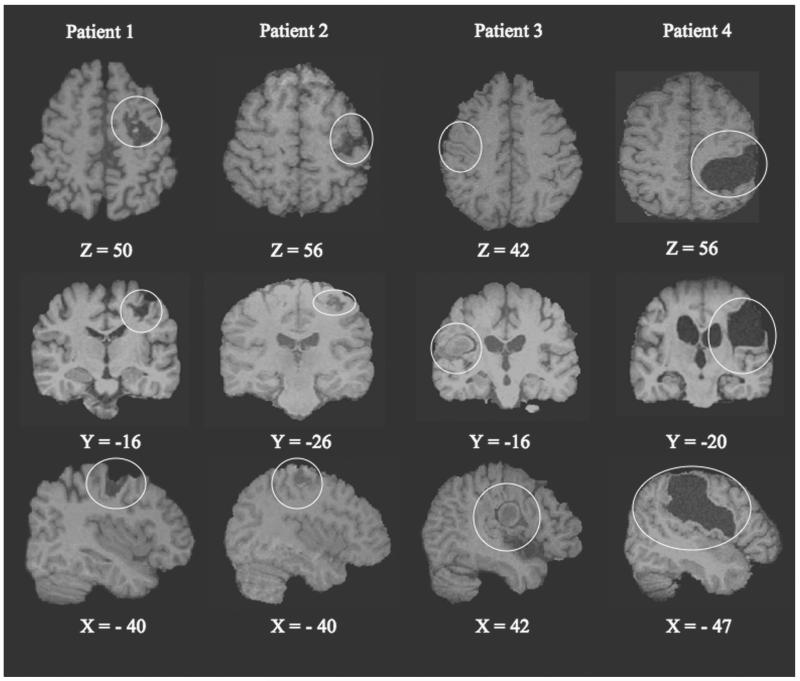
Figure 1.
Representative axial, coronal, and sagittal slices with Montreal Neurological Institute (MNI) coordinates illustrating the cortical lesion in relation to the primary motor cortex in each of the 4 patients with chronic hemiparesis (lesion outlined by fine circle). The z coordinates (mm) in MNI space that cover the hypointensity areas on T1-weighted magnetic resonance imaging (MRI) are 55 to 68 (patient 1), 62 to 69 (patient 2), 42 to 60 (patient 3), and 38 to 74 (patient 4). Slices are in radiological convention (right side of the brain to viewer’s left for top 2 rows).

