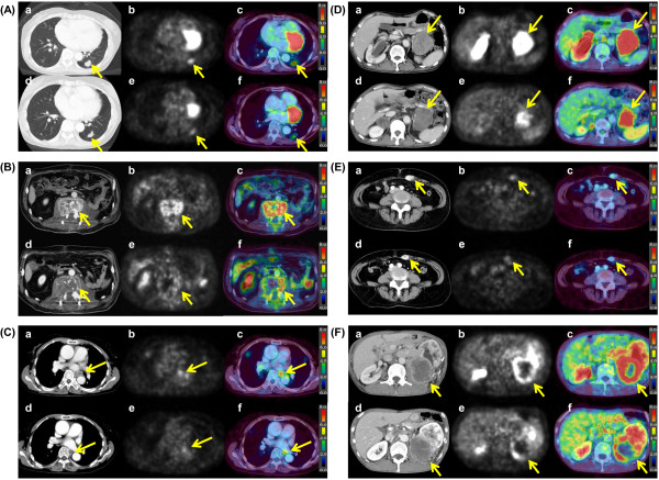
Figure 4.

FDG PET/CT features in the pretreatment state and post-treatment state. CT imaging (a), PET imaging (b) and fused imaging (c) before treatment and CT imaging (d), PET imaging (e) and fused imaging (f) at1 month after TKI treatment initiation. A: A 55-year-old female with lung metastasis. She was treated with sorafenib. The SUVmax showed a 25.6% decrease (from 4.3 to 3.2) and the tumor dia. showed a 13.6% decrease. B: A 66-year-old male with lumbar vertebrae metastasis. He was treated with sunitinib. The SUVmax showed a 27.5% decrease (from 8.0 to 5.8) and the tumor dia. showed a 3.9% decrease. C: A 76-year-old male with mediastinal lymph node metastasis. He was treated with sorafenib. The SUVmax showed a 13.2% decrease (5.3 to 4.6) and the dia. showed a 10.0% decrease. D: A 64-year-old male with adrenal gland metastasis. He was treated with sunitinib. The SUVmax showed a 25.0% decrease (8.8 to 6.6) and the dia. a 8.5% decrease. E: A 67-year-old female with rectus abdominis muscle metastasis. She was treated with sorafenib. The SUVmax showed a 26.5% decrease (3.4 to 2.5) and the dia. a 16.0% decrease. F: A 71-year-old male with primary tumor in the left kidney. He was treated with sunitinib. The SUVmax showed a 21.4% decrease (8.4 to 6.6) and the dia. did not change.
