FIGURE 4.
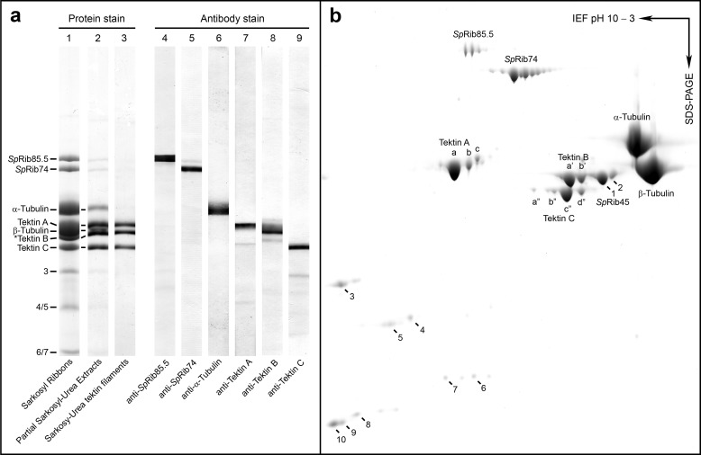
Composition of Ribbons, characterization of antibodies, and two-dimensional PAGE for MS analysis. a, lanes 1–3. SDS-PAGE protein staining of the following DMT fractions: lane 1, S. purpuratus Sarkosyl Ribbons (EM appearance shown in Fig. 2g) showing the principal constituent polypeptides; lane 2, ribbons partially extracted with urea to reduce tubulin that perturbs and obscures nearby bands; lane 3, Sarkosyl-urea-purified tektin filaments (Fig. 2h) composed of tektins A, B, and C. Masses as determined from their sequences are as follows: SpRib85.5, 85.5 kDa; SpRib74, 74 kDa; tektin A, 53 kDa; tektin B, 51 kDa; tektin C, 47 kDa; α- and β-tubulin, ∼50 kDa (*SpRib45 co-migrates with tektin B). Lanes 4–9 show immunoblots of S. purpuratus DMTs stained with the indicated antibodies. b, two-dimensional IEF/SDS-PAGE of Sarkosyl Ribbons. Major polypeptides (labeled) and spots 1–10 were reproducibly present and were cut out and analyzed by MALDI-TOF mass spectrometry (Table 2). Spots 1 and 2 were identified as SpRib45, homologue of Chlamydomonas CrRib43a. Corresponding positions of spots 3, 4/5, and 6/7 are indicated in a.

