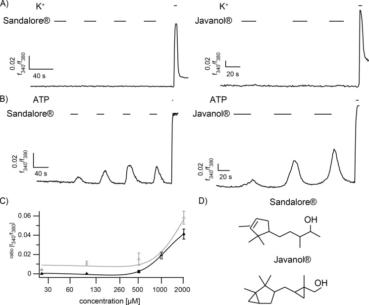
FIGURE 1.
A, representative calcium imaging experiments of monocultured trigeminal neurons that fail to react to Sandalore® and Javanol® stimulation (n = 413 and 85, respectively; 1 mm, 20 s). B, HaCaT keratinocytes in monoculture show repetitive calcium signals at each application of Sandalore® and Javanol® (n = 168 and 242, respectively; 1 mm, 20 s). ATP (100 μm) application served as a positive control. High potassium (K+; 45 mm) served as a positive control for neuronal cells. C, the dose-response curve of measurements of HaCaT cells after stimulation with 25, 100, 500, 1000, and 2000 μm Sandalore® (gray) or Javanol® (black). The mean amplitudes of stimulated HaCaT cells at the first application are shown. At 500 μm, the intracellular calcium was notably increased at the first application. D, the chemical structure of Sandalore® and Javanol®.

