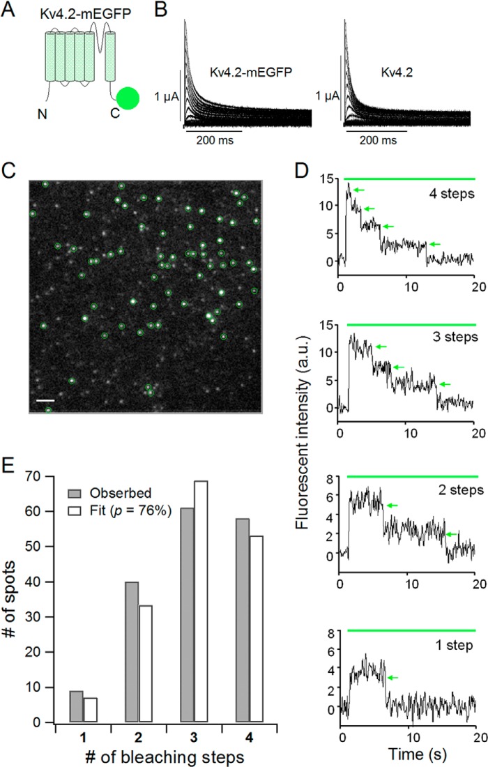
FIGURE 6.
Kv4.2 forms a tetramer. A, a schematic illustration of the Kv4.2-mEGFP construct. mEGFP was connected to the C terminus of Kv4.2 with a flexible GGS linker. B, currents from the Kv4.2-mEGFP construct (left) and WT Kv4.2 (right). The currents were elicited by step pulses from −100 to +50 mV in 10-mV increments. C, a single image from a TIRF movie of a Xenopus oocyte expressing Kv4.2-mEGFP. Green circles, countable spots for the bleaching step analysis. Scale bar, 2 μm. D, representative traces of 4, 3, 2, and 1 bleaching step(s) from Kv4.2-mEGFP. Green bar, illumination with 488 nm to excite mEGFP. Green arrows, bleaching events. a.u., arbitrary units. E, distributions of the number of spots with observed bleaching steps (gray) were fitted by a binomial distribution with p = 76% (white). p is the probability that the mEGFP attached to Kv4.2 is fluorescent.

