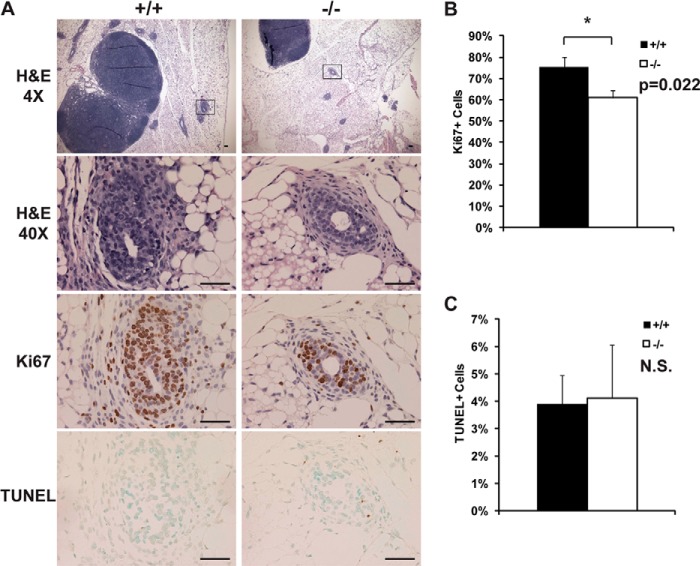
FIGURE 5.
Normal histology and apoptosis in Jarid1b−/− mammary glands are accompanied by increased cell proliferation. A, representative H&E, Ki67, and TUNEL images of Jarid1b+/+ and Jarid1b−/− mammary glands. Scale bar, 50 μm. B and C, quantification of Ki67+ (B) and TUNEL+ (C) cells. At least three pairs of female littermates were used per group, and three independent TEBs or proliferative ends were selected from different fields of view per mammary gland for quantification. Positively stained cells were counted by ImageJ. Error bars represent S.E. *, p < 0.05. N.S., not significant.

