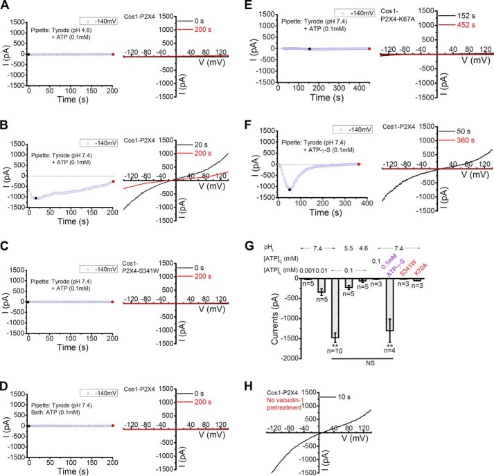
FIGURE 3.
Activation of lysosomal P2X4 by luminal ATP in a dose and pH-dependent manner. A, no current was detected in lysosomes from cells expressing rP2X4-GFP when a pipette solution (lysosome lumen) containing 0.1 mm ATP in pH 4.6 Tyrode was used. For this and subsequent figures, left, representative traces of current development at −140 mV; right, current-voltage relationships obtained by the voltage ramp at the time points indicated. B, large currents were detected with the use of a pipette solution containing 0.1 mm ATP in pH 7.4 Tyrode in lysosomes expressing rP2X4-GFP. C, no current was induced by 0.1 mm ATP in pH 7.4 Tyrode in lysosomes expressing rP2X4-S341W-GFP, a non-functional mutant. D, application of ATP (0.1 mm) in the bath (cytosolic side) failed to induce current in lysosomes expressing rP2X4-GFP. E, little current was induced by pipette infusion of pH 7.4 Tyrode containing 0.1 mm ATP in lysosomes from cells expressing rP2X4-K67A-GFP, an ATP binding deficient mutant. F, pipette infusion of 0.1 mm ATP-γS induced large currents in lysosomes expressing rP2X4-GFP. G, quantification of currents at −140 mV at conditions as indicated. Lysosomal P2X4 was activated by luminal, but not cytosolic, application of ATP or ATP-γS when the luminal pH was clamped at 7.4. ** (p < 0.01) indicates significant difference comparing with the currents induced by luminal 0.1 mm ATP at pH 4.6. H, large vacuoles obtained from rP2X4-GFP-transfected COS1 cells without the treatment of vacuolin-1 showed the same current-voltage relationship as the enlarged vacuoles induced by vacuolin-1.

