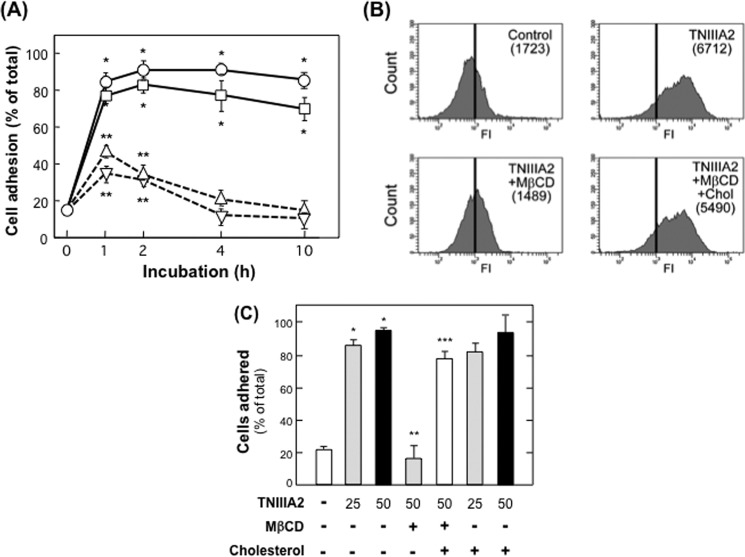
FIGURE 3.
Characterization of proadhesive nature of TNIIIA2. A, induced adhesion of K562 cells to the fibronectin by TNIIIA2. K562 cells were seeded on a 96-well plate coated with the fibronectin (10 μg/ml) in the presence of TNIIIA2 (25 μg/ml; circles), TS2/16 (20 μg/ml; squares), phorbol myristate acetate (50 ng/ml; triangles), or stem cell factor (50 ng/ml; inversed triangles). After culturing for the indicated time, cell adhesion to the fibronectin substrate was quantified as described previously (32, 33). Each point represents the mean ± S.D. of triplicate determinations. 1 of 3 individual experiments is shown. *, p < 0.005 versus untreated (0 h); **, p < 0.05 versus untreated (0 h). B, K562 cells were incubated with or without TNIIIA2 (25 μg/ml) in the presence or absence of Mβ-CD (10 mm) or Mβ-CD plus cholesterol (Chol; 16 mg/ml) inclusion complex for 30 min. Activation of β1-integrin was determined by flow cytometry using mAb 9EG7 recognizing the active β1 conformation specific epitope. C, to evaluate the effects on cell adhesion to the fibronectin substrate, K562 cells treated as above were seeded to 96-well plates coated with the fibronectin and incubated for 1 h. Cells adhering to fibronectin were fixed with formalin, and the numbers of cells were counted (C) as in Tanaka et al. (33). Data are representative of two individual experiments. *, p < 0.001 versus without TNIIIA2. **, p < 0.005 versus treated with TNIIIA2 alone. ***, p < 0.01 versus treated with TNIIIA2 and Mβ-CD.

