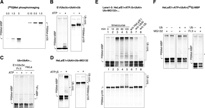
FIGURE 7.
A, cell-free T7 TnT expression of p105 and rhMBP. The indicated amount of reaction mixture (μl) was subjected to PAGE, dried, and visualized with a PhosphorImager. B–D, in vitro translated [35S]Met-labeled rhMBP and p105 were incubated with E1/Ubc5c supplemented with Ub and ubiquitin aldehyde with or without ATP (B); E1/Ubc5c supplemented with Ub, fraction II, and ubiquitin aldehyde with or without ATP (C, left); HeLa extract supplemented with Ub and ubiquitin aldehyde with or without ATP (C, right), and HeLa extract supplemented with E1, Ub, ubiquitin aldehyde, and MG132 in the presence or absence of ATPγS. E, in vitro-translated [35S]Met-labeled MBP (i) and p105 (ii) ubiquitinated in a cell-free system in the presence of HeLa extract supplemented by E1, ATPγS, ubiquitin aldehyde, Ub, and MG132. The reactions were terminated at the indicated time points and subjected to PAGE (lanes 1–5). Lanes 6–8 show the reactions performed for 120 min without the indicated components. F, degradation of in vitro translated [35S]Met-labeled rhMBP monitored in HeLa/E1-based cell-free systems supplemented with fraction II in the presence or absence of Ub and MG132, as indicated. WB, Western blotting; Ub conj., ubiquitin conjugates; Fr.II, fraction II; UbAl, ubiquitin aldehyde.

