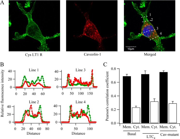
FIGURE 2.
Subcellular distribution of caveolin-1 and CysLT1 receptors in RBL-1 cells. Cells were co-transfected with caveolin-1-RFP and FLAG-tagged CysLT1 receptor and then fixed 48 h later. A, confocal images for the conditions shown. Line scans are shown in the merged panel. B, fluorescence profiles from the line scans are shown. Caveolin-1-RFP distribution is shown in red, and FLAG-tagged CysLT1 receptors are in green. C, histogram compares Pearson's correlation coefficient for the conditions shown. CaV-mutant denotes caveolin-1 with point mutations in the scaffolding domain (see Fig. 4). Mem., membrane; Cyt., cytosol.

