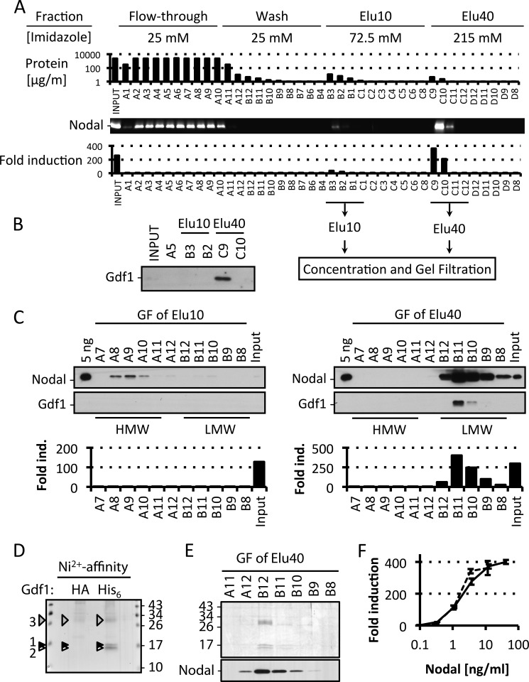
FIGURE 5.
Purification of Nodal·Gdf1 reveals active LMW complexes containing both prodomains. A, representative Nodal·Gdf1(His6) affinity purification profile from 45 ml of conditioned medium. Each 5-ml fraction was analyzed for total protein content by Bradford assay, Nodal protein content was analyzed by immunoblotting, and Nodal activity was analyzed in CAGA-luc reporter assays. B, immunoblot analysis of Gdf1 in relevant fractions. C, typical Nodal·Gdf1 gel filtration (GF) profile from pooled and concentrated Elu10 and Elu40 fractions from the affinity purification step. Each 5-ml fraction was analyzed by immunoblotting for Nodal and Gdf1 protein and by luciferase reporter assays for Nodal activity. D, silver staining of material eluted from a nickel affinity column using either HA- or His6-tagged Gdf1 in Nodal·Gdf1 conditioned medium. LC-MS/MS analysis identified mature Nodal and Gdf1 (black and white arrowheads, respectively) as well as the prodomains of Nodal and Gdf1 (gray arrowhead). E, selected fractions from the gel filtration of an Elu40 pool. Top, silver staining. Bottom, immunoblotting against Nodal. F, induction of CAGA-luc by affinity-purified (dashed line) or gel-filtered (solid line) Nodal·Gdf1. In F, error bars represent S.D. ind., induction.

