
FIGURE 6.
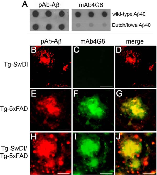
Analysis of non-mutated Aβ and Dutch/Iowa CAA mutant Aβ accumulation in Tg-SwDI, Tg-5xFAD, and bigenic Tg-SwDI/Tg-5xFAD mouse brains. A, a dot blot analysis was performed to demonstrate that rabbit polyclonal antibody to human Aβ (pAb-Aβ) recognizes both non-mutated Aβ and Dutch/Iowa CAA mutant Aβ, whereas mAb4G8 only recognizes non-mutated Aβ. Brain sections obtained from the different transgenic mouse lines at 9 months of age were immunolabeled with pAb-Aβ (red, B, E, and H), mAb4G8 (green, C, F, and I), and merged (D, G, and J). Dutch/Iowa CAA mutant Aβ deposits in Tg-SwDI mice are immunolabeled with pAb-Aβ (B) but not mAb4G8 (C). Non-mutated Aβ deposits were immunolabeled with both pAb-Aβ (E) and mAb4G8 (F). Aβ plaque core deposits in bigenic Tg-SwDI/Tg-5xFAD mice were immunolabeled with both pAb-Aβ (H) and mAb4G8 (I). The merged image shows a halo of Aβ deposits around the periphery of plaques that were not immunolabeled with mAb4G8, suggesting that they are composed of Dutch/Iowa CAA mutant Aβ (J). Scale bars = 50 μm.