
Fig. 3.
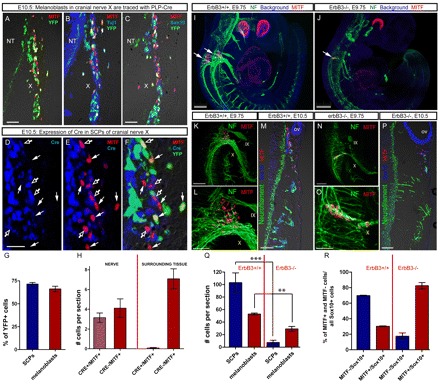
Genetic tracing using PLP-CreERT/Rosa26-YFP mice and analysis of neuregulin receptor subunit (Erbb3) mutant mice reveal SCP-derived melanoblasts in cranial nerves IX-X. (A-C) Injection of TM at E9.5 and analysis at E10.5 of Mitf and YFP (A); Mitf, Tuj1, YFP (B); and Mitf, Sox10, YFP (C). Note the presence of YFP in a significant proportion of Mitf+ cells in the Tuj1+ and Sox10+ nerve and in Mitf+ cells associated with but not within the nerve. (D-F) Immunohistochemical staining for Cre recombinase, Mitf and YFP in PLP-CreERT2 mice at E10.5. Note the expression of Cre in SCPs of nerves, including those that are weakly Mitf+ within nerves (unfilled arrows), but not in strong Mitf+ cells (filled arrows). (G) Quantification of YFP+ cells in Sox10+ SCPs of nerves and in Mitf+ melanoblasts (n=4). (H) Quantification of Cre expression in Mitf+ cells in SCPs of nerves and melanoblasts outside cranial nerves IX-X. (n=4). (I,J) Whole-mount immunohistochemistry of E9.75 wild-type (I) and ErbB3–/– (J) mouse embryos stained for NF (green) and Mitf (red). Arrows indicate clusters of melanoblasts associated with cranial nerves IX-X. (K,L) Cranial nerves IX-X of an E9.75 ErbB3+/+ embryo. (M) Cross-section through cranial nerves IX-X of an E10.5 ErbB3+/+ embryo. Note the numerous Sox10+ SCPs and Mitf+ melanoblasts associated with NF+ nerve fibers. (N,O) Cranial nerves IX-X of an E9.75 ErbB3–/– embryo. (P) Cross-section through cranial nerves IX-X of an E10.5 ErbB3–/– embryo. Note the reduction of Sox10+ SCPs and Mitf+ melanoblasts. (Q) Cell numbers of SCPs and melanocytes in wild-type and ErbB3–/– E10.5 embryos (***P=0.001, n=4 embryos/genotype). (R) Percentage of melanoblasts (Sox10+:Mitf+) and SCPs (Sox10+:Mitf–) relative to all Sox10+ cells in wild-type and ErbB3–/– E10.5 embryos. Note that the proportion of Mitf+ cells is much higher in ErbB3–/– than in wild-type embryos. Error bars represent s.e.m. Red line in H, Q and R separates analyzed tissues or genotypes. NT, neural tube; ov, otic vesicle. Roman numerals indicate the cranial nerves. Scale bars: (A-C) 50 μm inA-C; 30 μm in D-F; 250 μm in I,J; 100 μm in K,M,N,P; 50 μm in L,O.