
Fig. 6.
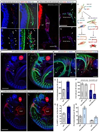
Sox2 regulatory effects on melanocyte development in NCC conditionally mutant mice (Wnt1Cre/Sox2fl/fl). (A-D) Expression of Sox2 (blue) and Mitf (red) in cells associated with cranial nerve IX in E10.5 mouse embryos. White arrows point at Mitf+ cells with no or very low levels of Sox2 (C,D), yellow arrows show Mitf+ cells retaining low levels of Sox2 (C,D). (E) Sox2 expression (red) decreases proximo-distally in the ventral spinal nerve (SN) in the E12 mouse embryo. (F,G) Sox2 expression in SCPs of spinal nerve dorsal rami. Arrows indicate Sox10+ prospective melanoblasts under the epidermis that are negative for Sox2. (H) Schematic model of Sox2 and Mitf cross-repressive interactions during development of the glial and melanocyte lineages. (I-L) Conditional deletion of Sox2 in the NCCs using the Wnt1-Cre activator strain. Melanoblasts numbers at the cranial nerves IX-X cluster markedly increased in conditional null mice (K,L) compared with control mice (I,J). (M,N) Magnified view of part of J and L. (O) Quantification of Mitf+ cells in IX-X cluster of wild-type and Wnt1Cre/Sox2fl/fl mice at E10.5 (left graph, P<0.0001, n=4). (P) Quantification of all Sox10+ cells and Sox10+/Mitf– cells in cranial nerves IX/X of Wnt1Cre/Sox2fl/fl and wild-type embryos (for Sox10+/Mitf– cells **P=0.0096, n=4 embryos/genotype). (Q) Percentage of Mitf+/Sox10+ cells among all Sox10+ cells between Wnt1Cre/Sox2fl/fl embryos and littermate wild-type embryos (**P=0.0012, n=4 embryos/genotype). (R) Quantification of melanoblasts appearing adjacent to cranial ganglia V and VII/VIII in Wnt1Cre/Sox2fl/fl embryos and littermate control embryos (ganglion V: *P=0.0161, n=3 Wnt1Cre/Sox2fl/fl and n=5 in control; ganglia VII/VIII: **P=0.0014, n=4 Wnt1Cre/Sox2fl/fl and n=8 in control). (O-R) Error bars represent s.e.m. NT, neural tube; ov, otic vesicle; SN, ventral spinal nerve. Roman numerals indicate the cranial nerves. Scale bars: 100 μm in A,B,F,G,M-P; 50 μm in C,D; 500 μm in I-L.