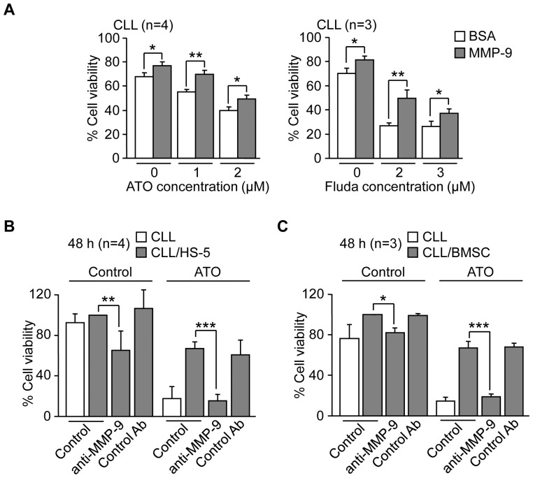
Figure 6. MMP-9, isolated or present in stroma, induces resistance of CLL cells to ATO and fludarabine.
(A) 1.5×10⁵ CLL cells RPMI/0.1% FBS were cultured on 0.5% BSA or 150 nM MMP-9 for 1 h prior to adding the indicated concentrations of ATO or fludarabine (Fluda). After 24 h (ATO) or 48 h (Fluda) cell viability was determined by flow cytometry using FITC-Annexin V and PI. (B) 1.5×10⁵ CLL cells were treated or not with anti-MMP-9 pAbs or control pAbs for 1 h and added to wells coated with 0.5% BSA, HS-5 cells or primary stromal cells (BMSC). After 2 h at 37°C, 2 µM ATO was added and cells further incubated for 48 h. Cell viability was determined by flow cytometry using FITC-Annexin V and PI. The viability of CLL cells cultured over stroma in the absence of ATO was normalized to 100 and average values are shown. *P≤0.05; **P≤0.01; ***P≤0.001.

