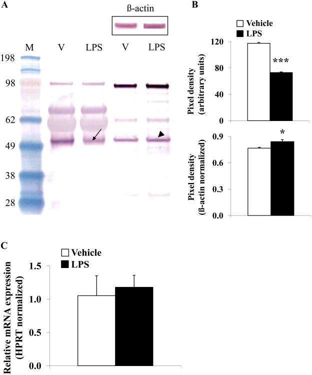
Fig. 2.
LPS decreases plasma GOAT concentration, whereas protein content in the gastric corpus is slightly increased and mRNA expression unchanged in rats. Overnight fasted rats were injected ip with LPS (100μg/kg body weight) or vehicle (pyrogen-free saline) and trunk blood and stomach were collected at 2h post injection. Equal amounts of protein were loaded and plasma and gastric corpus mucosa GOAT concentrations were assessed using Western blot followed by semi-quantitative analysis. Lane 1 contains the molecular weight standards, lane 2 plasma proteins after vehicle injection, lane 3 plasma proteins after LPS, lane 4 gastric corpus mucosa proteins after vehicle and lane 5 gastric corpus mucosa proteins after LPS (A). The blot shows two dominant bands at ~50 and ~100 kDa. The 50 kDa band represents monomeric GOAT, whereas the 100 kDa band likely represents an SDS-stable dimer. Injection of LPS reduced the 50 kDa band (arrow) compared to vehicle demonstrating reduced plasma concentration of GOAT, whereas GOAT in the gastric corpus mucosa was increased (arrowhead, A). Re-staining of the Western blot with β-actin demonstrates equal gastric corpus mucosal protein concentration (A, insert). Quantification of GOAT plasma and stomach protein expression is shown in (B). Gastric GOAT mRNA expression did not change at 2h post LPS injection compared to vehicle treated rats (C). * p < 0.05 and *** p < 0.001 vs. vehicle; LPS, lipopolysaccharide; M, standard molecular weight marker; V, vehicle.

