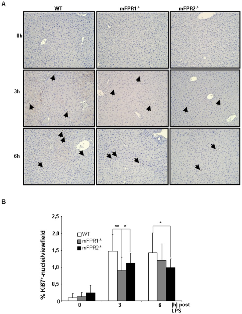
Figure 7. To investigate liver proliferation FFPE-sections were stained with the universal cell cycle marker Ki67.
At 3+-nuclei were counted and analyzed as percentage of proliferative cells. Photomicrographs were taken at 200-fold and representative images are shown. Ki67+-nuclei are indicated by arrows (* = p<0.05; ** = p<0.01).

