
Figure 1. Bronchointerstitial pneumonia in pigs infected with influenza A virus.
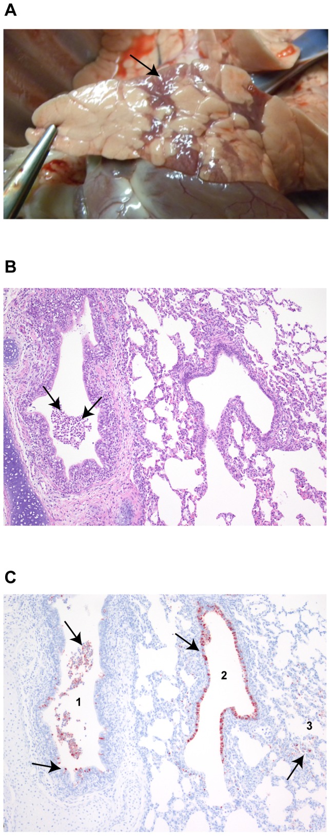
Pathological changes were recorded in the lungs of twelve pigs inoculated with influenza A virus on days 1–5 and 8 pi (n = 2 per day). (A) Macroscopic lesions were most prominent on day 3 pi; representative lesions are shown (arrow). (B) Histological changes on day 1 pi; representative lesions are shown. Leucocytes and cell debris (arrows) in the lumen of a bronchus. Hematoxylin and eosin, 100x. (C) Influenza A virus NP positive staining (arrows) in luminal cells and in the epithelial lining of a bronchus (1), a bronchiole (2) and in the pneumocytes of the parenchyma (3) in the same animal as shown in (C), serial sections (NP stained with AEC and counterstained with hematoxylin, 100x).