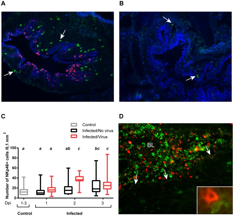
Figure 4. NKp46+ cells in the lungs of influenza virus infected pigs.
Lung tissue sections from pigs infected with influenza A virus and control pigs were stained with immunofluorescence markers for cytokeratin (blue), NKp46 (green) and influenza A virus NP (red). NKp46+ cells were counted in areas were influenza A virus NP was (A) detected and (B) not detected. Representative pictures taken from the same animal on day 1 pi are shown. Arrows point at NKp46+ cells. Immunofluorescence staining, 200x. (C) Plot shows number of NKp46+ cells per 0,1 mm2 in sections (n = 24 per animal) from control animals (n = 6) and in areas with and without virus in infected animals (n = 4 per day) calculated as described in Material and Methods. Groups with different letters differ significantly (p≤0.05). (D) NKp46+ cells in the lumen of a bronchus (BL). Arrows point at the epithelial lining. Representative picture of luminal exudate, taken from an infected animal on day 2 pi. Insert shows NKp46+ and influenza A virus NP+ cell in the lung tissue of an infected animal on day 1 pi. Immunofluorescence staining, 400x.

