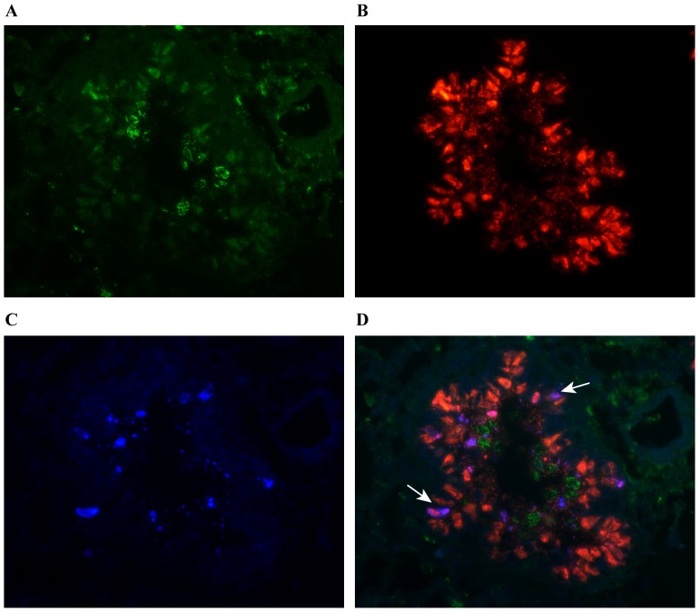
Figure 5. Staining for apoptosis in the lungs.
Lung tissue sections from animals infected with influenza A virus (n = 12) were stained with immunofluorescence markers against (A) NKp46(green), (B) influenza A NP(red) and (C) the apoptosis marker caspase-3(blue). (D) Overlay displaying simultaneously influenza A virus NP+ and caspase-3+ cells as purple (arrows). Representative of virus infected bronchiole at day 1 pi. Immunofluorescence staining, 400x.

