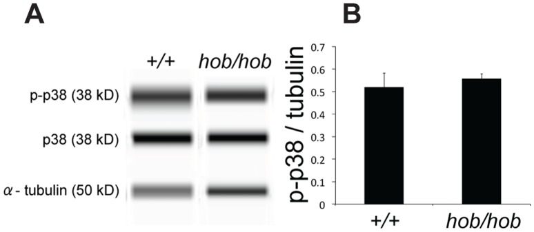
Figure 5. Quantitation of phospho-p38 MAPK (p-p38) levels in gonadal samples at 11.5 dpc (16–18 ts) in XY wild-type and Fgfr2hob/hob gonads.

A) Lane view images showing Simple Western detection of p-p38, p38, and α-tubulin. B) Graph showing the ratio of p-p38 to tubulin in the two gonadal genotypes. The ratio of p-p38 to p38 was similarly unaltered. Errors were calculated using standard error mean.
