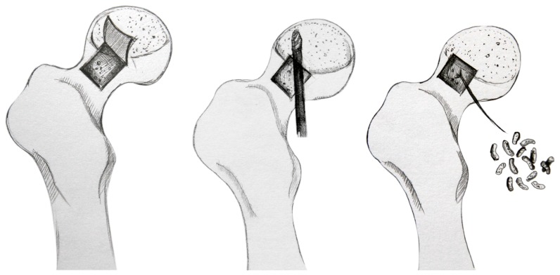
Figure 1. Schematic diagram of “Light Bulb procedure”: An approximate 1.5 cm×1.5 cm bone window was made at the femoral head–neck junction using osteotomes.
And the necrotized bone located at anterolateral and upper side of the operated femoral head was alternately debrided using a high-speed drill. Then the cavity was filled with an autologous cancellous bone combination of rhBMP-2.

