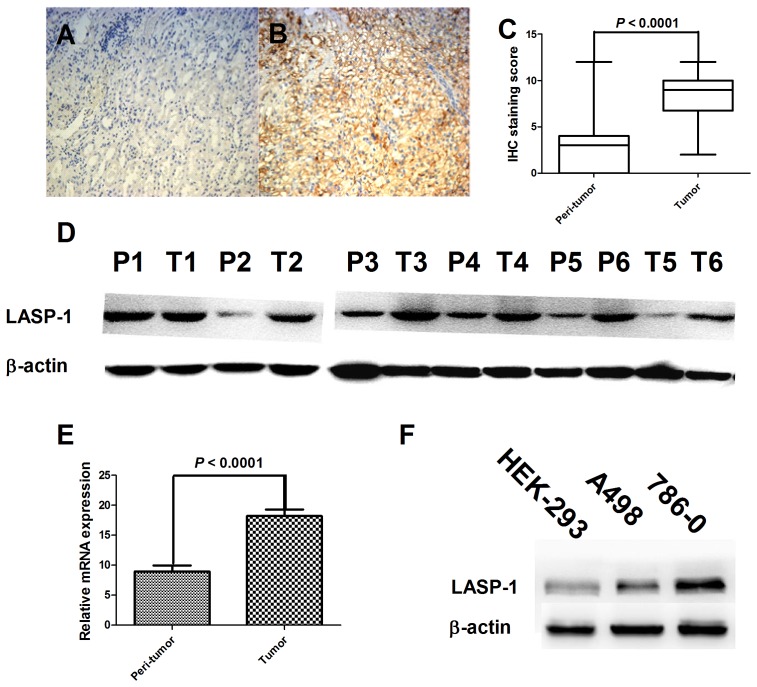
Figure 1. LIM and SH3 protein 1 (LASP-1) expression in clear cell renal cell cancer (ccRCC) tissues and cell lines.
LASP-1 protein expression in paraffin-embedded ccRCC tissues (A) and adjacent nontumorous tissues (B) using immunohistochemistry (magnification, 100×), in which positive LASP-1 immunostaining showed brown color. Wilcoxon analysis demonstrated that tumor tissues showed significantly higher LASP-1 expression than nontumorous tissues (C, n = 216). Western blot (D) and real-time PCR (E, n = 20) analyses confirmed the findings in immunohistochemistry analysis. Western blot analysis also showed differential LASP-1 expression in human embrynal kidney cells (HEK-293) and ccRCC cell lines (F). T refers to tumor tissues, whereas P refers to peritumor (nontumorous) tissues in panel D.

