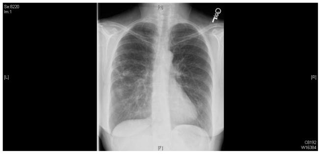
Image 1.

The chest X-ray shows diffuse interstitial fibrotic-type opacities throughout both lungs. There are ill-defined somewhat nodular appearing densities with a hint of cavitation in them on the right side, particularly in the apical segment of the lower lobe. Mild scoliosis is notable.
