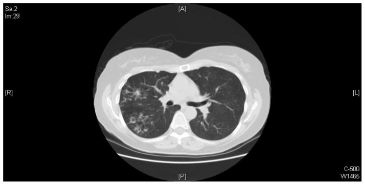
Image 3.

The representative image of chest CT scan shows that multiple small airways opacities are scattered in the right upper lobe, superior right lower lobe, and right middle lobe. Small areas of bronchiectasis are present in the right upper lobe. The largest area of cystic bronchiectasis/small cavity formation measures approximately 1 centimeter and it is present in the posterior right upper lobe.
