Figure 3.
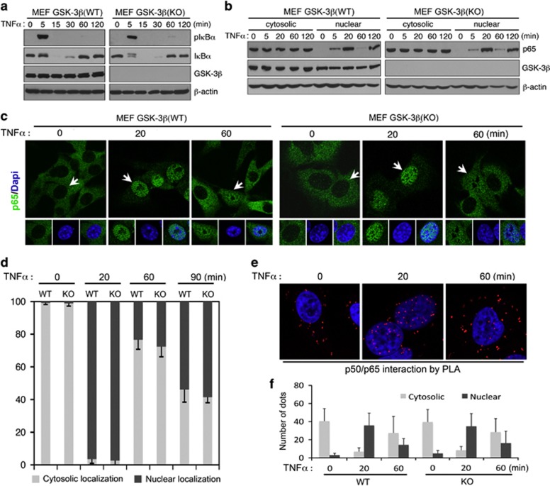
GSK-3β is not essential for TNFα-induced IKBα degradation or p65 nuclear translocation. (a, b) Western blot analysis of whole-cell extracts (a) or cytosolic and nuclear extracts (b) from MEFs treated with 10 ng/ml TNFα for the indicated periods of time. Note that there is no discernible difference in TNFα- induced IκBα phosphorylation/degradation (a) or p65 nuclear translocation (b) between WT and GSK-3β-null MEF cells. (c) Immunofluorescence staining and confocal microscopic imaging showing p65 expression/localization in MEF cells following TNFα stimulation (10 ng/ml) for the indicated periods of time. (d) Quantification of images (c) for percentage of p65 with predominant cytoplasmic or nuclear localization in wild-type (WT) and Gsk-3β−/− (KO) MEF cells at the indicated time points. Arrows point to the cells shown in inset with p65 predominantly localized to the cytoplasm (0 min) or nucleus (20 and 60 min). (e) Confocal microscopic imaging showing in situ p50 and p65 interaction detected by PLA following TNFα stimulation. (f) Quantification of dots corresponding to sites of interaction as shown in (e) as mean±S.D. as described in Materials and Methods

