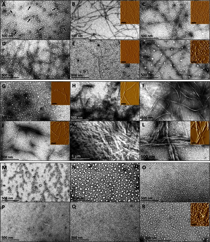
Fig. 3.
TEM and AFM micrographs of peptides DRV(9–24), KLL(45–63), and VLES(221–239). Evolution of peptide aggregation in water was evaluated by electron and atomic force microscopies on peptides VLES(221–239) (A–F), DRV(9–24) (G–L), and KLL(45–63) (M–R). Incubation times correspond to: 0 h (A, G, M), 24 h (B, H, N), 48 h (C, I, O), 72 h (D, J, P), 92 h (E, K, Q), and 120 days (F, L, R). Arrows (A) show protofibrillar structures. Insets correspond to AFM images of samples used in CD experiments and the scale correspond to 1 µm

