
Figure 1. Slc25a12-knockout mice have smaller brains.
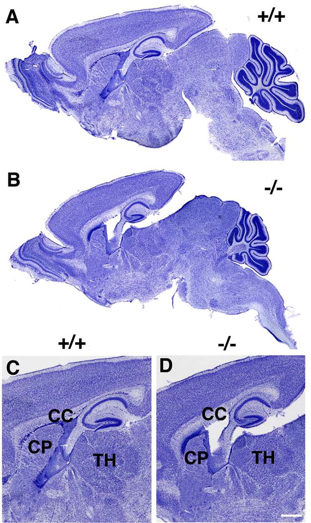
Sagittal sections of P13 brain from wild type (+/+, A, C) or knockout (−/−, B, D) mice are shown stained with Cresyl violet. Note the generally smaller size of all brain regions in the knockout animal. Panels C and D show higher power views of the images in A and B, with the corpus callosum (CC), caudate putamen (CP) and thalamus (TH) indicated. Scale bar: A and B, 750 μm; C and D, 500 μm.