
Figure 2. Slc25a12-knockout mice show myelination deficits.
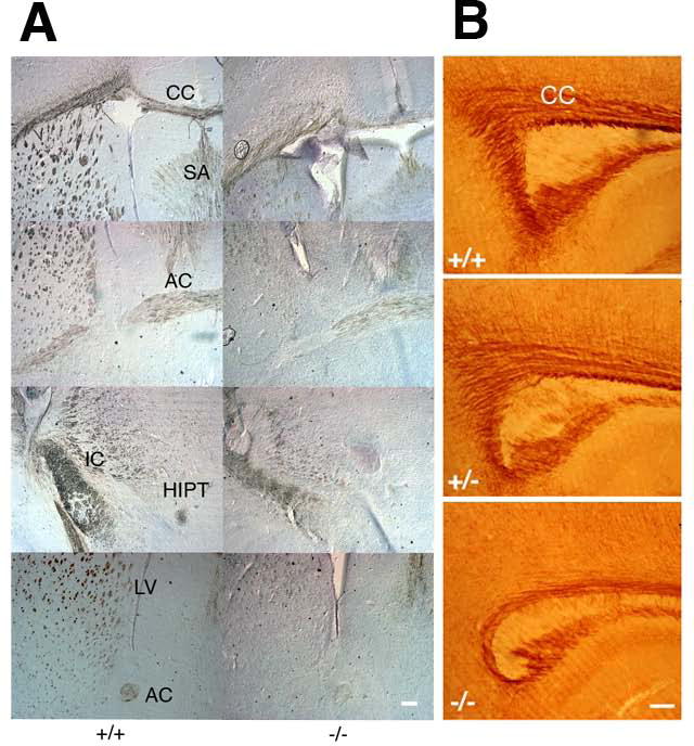
A. Coronal sections through frontal cortex from P14 wild type (+/+) and knockout (−/−) brains were stained with anti-MBP antibody followed by HRP-conjugated secondary antibody visualized by the DAB reaction. B. Sagittal sections from P13 wild type (+/+), heterozygous (+/−) and knockout (−/−) brains were stained with an anti-PLP antibody. CC, corpus callosum; SA, septal area; IC, internal capsule; AC, anterior commissure; HIPT, habenulo-interpedunclar tract; LV, lateral ventricles. Scale bars, 200 μm.