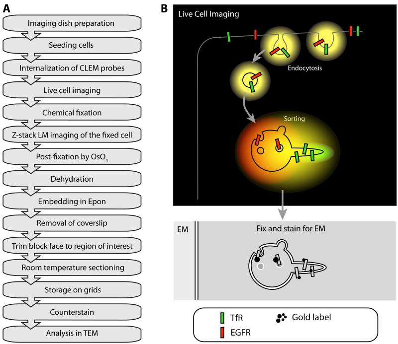
Figure 4. The principle of CLEM.
A: Flow diagram of a typical CLEM experiment using the “simple” approach described in III.3.c consisting of live labeling, chemical fixation and Epon embedding. (OsO4) osmium tetroxide B: Cartoon of a CLEM experiment on RFP-EGFR and GFP-TfR trafficking. Both types of cargo are internalized by endocytosis, showing up as yellow fluorescent compartments in live cell imaging due to the co-localization of GFP and RFP. In the early endosome, GFP-TfR is sorted into tubular/vesicular carriers that retrieve the receptor to plasma membrane. In the fluorescence microscope, these carriers appear green due to the high concentration of GFP-TfR. The RFP-EGFR is collected in intraluminal vesicles in the endosomal vacuole, which shows up as predominately red. Although this sorting process is visible at the LM level, the endosome itself or its subdomains are not visible. By capturing the event by HPF and analyzing the sample by EM, it is possible to define the localization of GFP-TfR and RFP-EGFR in their different subdomains within the early endosome compartment.

