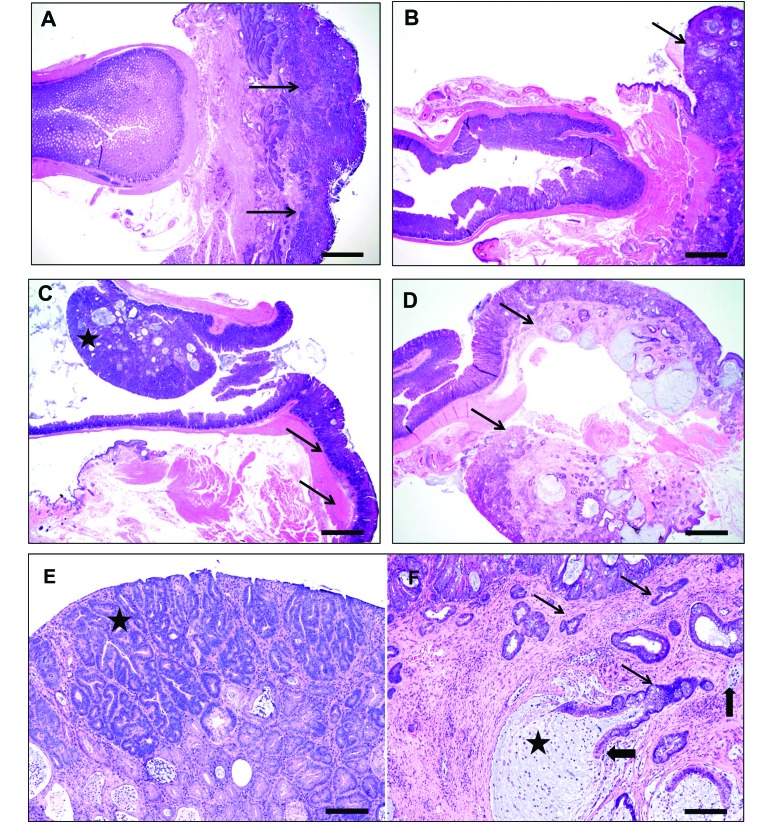
Figure 3.
Representative histology from categories of RP mice. (A) Distal gastrointestinal tract of an IL10−/− mouse, with prolapsed rectum (arrows) showing severe mucosal inflammation, ulcerations with marked glandular hyperproliferation, and dysplasia. Note the patchy multifocal inflammatory aggregates in the nonprolapsed distal colon. (B) Distal colon and prolapsed colorectum of a mouse from the immunocompromised group, showing prominent inflammation, glandular hyperplasia, distortion, and mucin-filled cystic glands in the prolapsed segment (arrow) and milder changes in the nonprolapsed distal colon. (C) Rectum of a mouse in the miscellaneous group, showing a distal colonic polypoid adenoma (star) and associated prolapsed segment (arrow) with inflammation, epithelial tethering, and hyperplasia. (D) Rectal prolapse (with junctional distal colon) from an Lpd−/− mouse, showing mucosal erosions, inflammation, and prominent hyperplastic, dysplastic and mucous-filled cystically distended invasive glands (arrow). Note that the lesions are mostly within the prolapsed segment and that the adjacent nonprolapsed colon is relatively unaffected. (E) Higher magnification of the mouse in panel C, showing darker staining adenomatous tubules (star) in superficial half arising from a background of hyperplastic, dysplastic, and cystic tubules with moderate stromal inflammation. (F) Higher magnification of the mouse in panel D, showing multiple irregular angular horizontally spreading proliferating dysplastic glands (thin arrows) deep in the submucos and musculature with nuclear atypia and piling and budding in a thickened stroma by fibrosis and inflammation. Frequently, the invasive cystic glands are filled with mucous (star) and have either partial or complete cell loss (thick arrows) from the lining epithelium and intaluminal cellular debris. Hematoxylin and eosin stain; bar, 400 µm (A through D); 80 µm (E and F).

