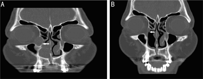
Figure 2. Pictures of pre-op and post-op of computed tomography.
A: Sinus CT coronal view (the patient): Inverted papilloma widening right side nasolacrimal sac and duct and protruding into right side nasal cavity; B: Sinus CT coronal view (the patient): There was no recurrence sign or residual tumor at right side nasolacrimal sac and nasal cavity after a three-year follow-up.

