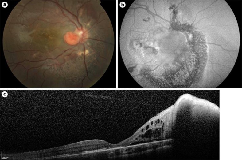
Fig. 2.
a Postoperative fundus examination showed a reduction in size of the JRCH, and the optic nerve head appeared almost completely free from the lesion. The JRCH was stable in size for the whole duration of follow-up. b Twenty-four months postoperatively, FAF imaging revealed a hypoautofluorescent round area dislocated from the optic disc and surrounded by a hypoautofluorescent area. c At the last follow-up, SD-OCT showed resolution of the macular exudation and reduction of papillomacular area fluid. The patient's BCVA had improved to 20/25.

