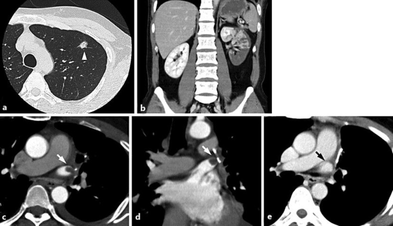
Fig. 1.
a Preoperative CT of the chest reveals a nodular shadow in the upper lobe of the left lung (arrowhead). b Enhanced CT reveals a large wedge-shaped defect in the left kidney. c, d Enhanced CT after the diagnosis of renal infarction reveals a round defect in the stump of the left superior pulmonary vein (white arrow). e Enhanced CT 2 months after renal infarction reveals resolution of thrombosis (black arrow).

