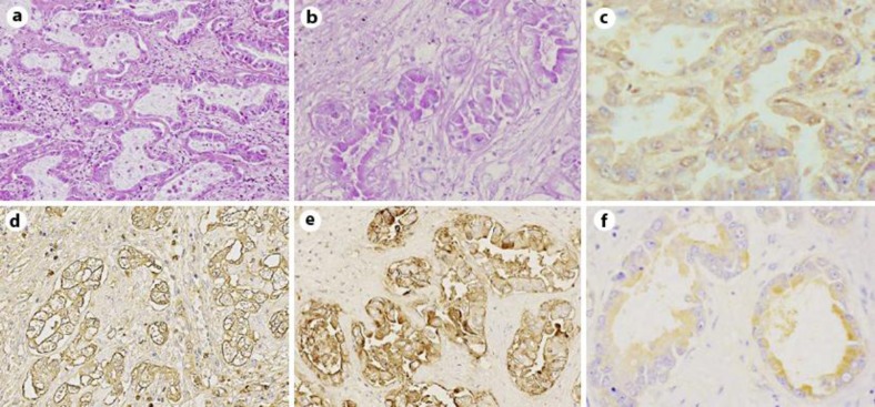
Fig. 3.
Intrahepatic cholangiocarcinoma. a HE, ×100. b PAS, ×200. c–f Immunohistochemistry for TF (c), MUC-1/Y (d), MUC-1 (e) and MUC-16 (f), respectively, ×200. TF and MUC-16 were moderately expressed in the cytoplasm of cholangiocarcinoma cells. MUC-1/Y and MUC-1 were expressed strongly in the cytoplasm of cholangiocarcinoma cells.

