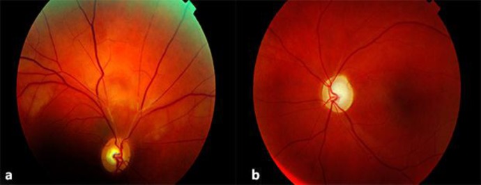
Fig. 1.

a Right fundus. A normal optic nerve with segmental obstructions of the arteries in the temporal and nasal superior branches. A pale ischemic edema is present in the retina superior to the optic nerve. b Left fundus. A pale atrophic optic nervehead and thin white ‘ghost vessels’ were observed in the superior temporal arcade.
