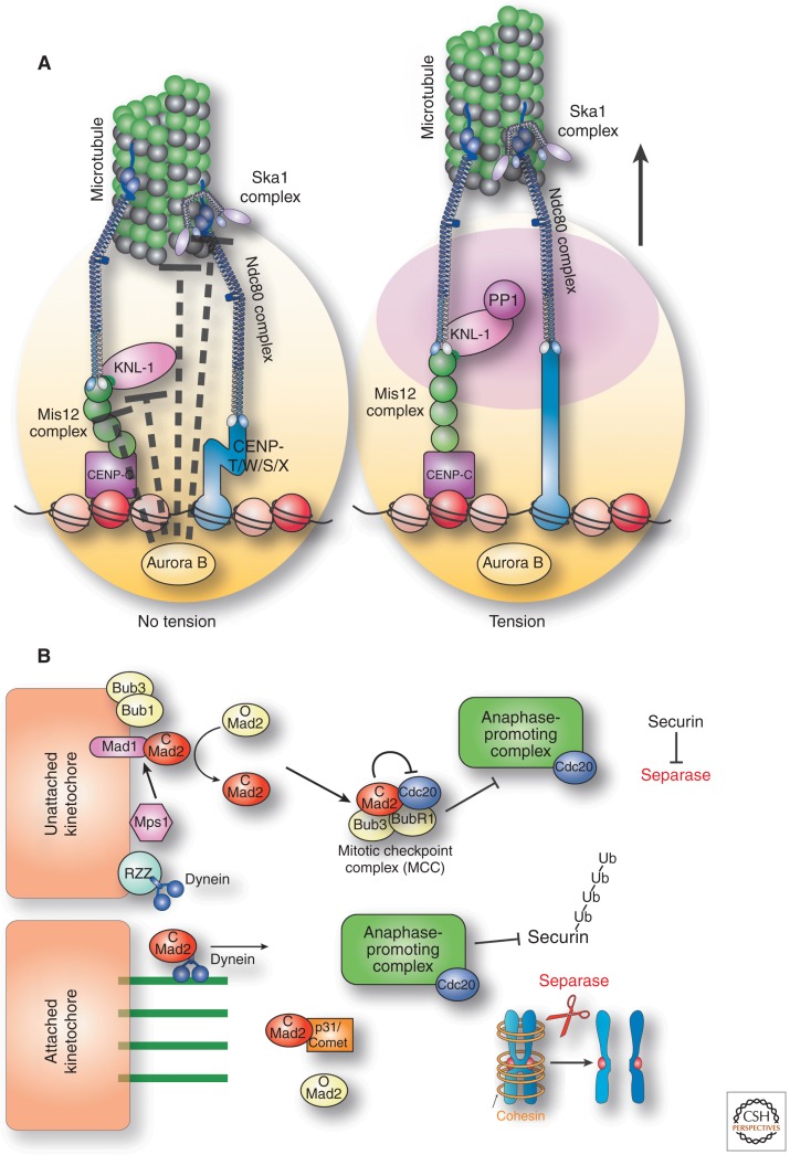
Figure 5.
Regulation. (A) Diagram showing the tension-dependent deformation of the kinetochore structure. Aurora B kinase located at the inner centromere (at the base of the kinetochore) is spatially separated from its substrates at the outer kinetochore. The presence of tension on bi-oriented kinetochores strongly reduces the ability of Aurora B to phosphorylate outer kinetochore proteins and inactivate microtubule attachments. (B) Model showing the spindle assembly checkpoint proteins preventing cell cycle progression in the absence of unattached kinetochores.

