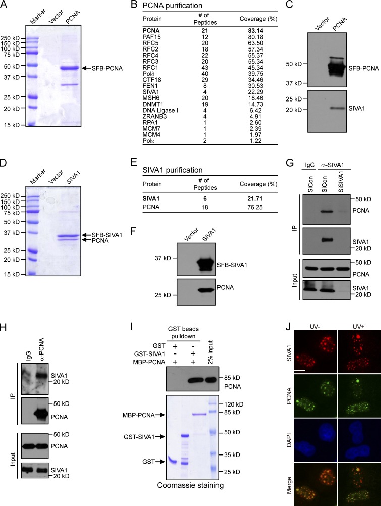
Figure 1.
Identification of SIVA1 as PCNA-binding partner. (A and D) HEK293T cells stably expressing either the vector control or SFB-tagged (S tag, Flag epitope tag, and streptavidin-binding peptide tag) PCNA/SIVA1 were used for tandem affinity purification of protein complexes. The final eluates were resolved on SDS-PAGE gels and Coomassie stained. (B and E) Tables are summaries of proteins identified by mass spectrometry analysis. Letters in bold indicate the bait proteins. (C and F) Confirmation of PCNA- or SIVA1-interacting proteins by Western blotting. PCNA/SIVA1 purification products were analyzed by Western blots using specific antibodies against Flag, SIVA1, or PCNA. (G and H) Association of endogenous SIVA1 with PCNA in HeLa cells was performed by coimmunoprecipitation using the anti-SIVA1 or anti-PCNA antibody. SiRNA-treated HeLa cells were lysed in the presence of Benzonase, cell lysates were then incubated with protein A agarose beads conjugated with the indicated antibodies, and Western blot analysis was performed according to standard procedures. siCon, control siRNA. (I) Direct in vitro binding between recombinant MBP-PCNA and GST-SIVA1 purified from E. coli. GST served as negative control for PCNA binding. (top) PCNA was detected by immunoblotting. (bottom) Purified proteins visualized by Coomassie staining. (J) SIVA1 colocalizes with PCNA at nucleus foci. HeLa cells were transfected with HA-Flag–tagged SIVA1 plasmids. 24 h after transfection, cells were treated with 50 J/m2 UV or left untreated. After 1 h recovery, foci assembled by this fusion protein and by PCNA were detected by immunofluorescence using anti-Flag and anti-PCNA antibodies, respectively. HA-Flag–SIVA1 foci were detected in red, whereas PCNA foci were detected in green. A merged image shows colocalization. IP, immunoprecipitation. Bar, 10 µm.

