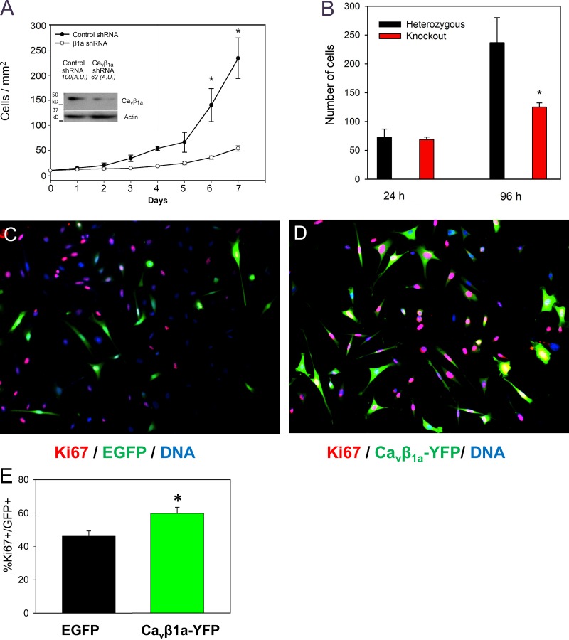
Figure 4.
Regulation of myoblast proliferation by Cavβ1a in vitro and in vivo. (A) Quantification of myoblast growth for 7 d after transfection with either scrambled control shRNA or Cavβ1a-targeted shRNA (Western blot of Cavβ1a knockdown is inset). (B) Quantification of MPCs cultured from Cacnb1+/− and Cacnb1−/− embryos for 4 d (n = 4). (C–E) Primary mouse myoblasts transfected with EGFP (C) or Cavβ1a-YFP (D) and stained 24 h later for Ki67 (red; n = 3). Bar, 100 µm. (E) Quantification of Ki67+/EGFP and Ki67+/Cavβ1a-YFP cells expressed as a percentage of total EGFP or Cavβ1a-YFP + cells. *, P < 0.05.

