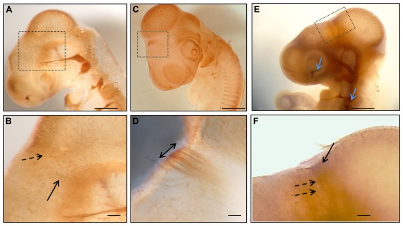
FIGURE 1.
Whole mount immunohistochemistry during the first stages of posterior commissure development. (A,B) HH17 embryos immunolabeled with 3A10 clone (DSHB) showing axons from neurons of the dorsal area of prosomere 1 (discontinuous arrow in B) and from the basal diencephalon (continuous arrow in B). (C,D) HH18 chick embryo immunostained with anti-tubulin βIII showing the posterior commissure as the first transversal tract on the dorsal encephalon (double arrow in D). (E,F) HH17 chick embryo immunolabeled with anti-tubulin βIII (brown) and anti-EphA7 (blue) showing the expression of EphA7 (continuous arrow in F) in the dorsal diencephalon concomitantly with the axons of the posterior commissure (discontinuous arrows in F), as well as in control points such as the retina and otic vesicle (blue arrows in E). (B,D,E) Magnification of the inset in (A,C,E), respectively. Bars in (A,C,E) = 0.5 mm; and in (B,D,F) = 0.1 mm.

