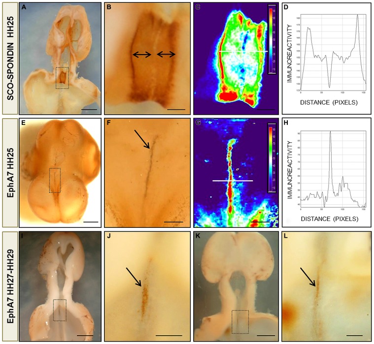
FIGURE 4.
Protein pattern expression of EphA7 and SCO-spondin during chick brain development. (A,B,E,F) Whole mount immunohistochemistry for SCO-spondin (A,B) and EphA7 (E,F) of HH25 chick brain embryos showing the complementarity expression between the labeling of SCO-spondin and EphA7 in the diencephalic roof plate (arrows in B,F). (C,G) Pseudocolor images of (B,F) showing the expression level of SCO-spondin and EphA7, respectively. The intensity of the immunoreaction of the white line in (C,G) was analyzed in (D,H). (I,J) and (K,L) Whole mount immunohistochemistry for EphA7 in the diencephalic roof plate in later stages of development (HH27 and HH29, respectively) showing that the expression of EphA7 continues at latter stages but with a lower intensity (arrows in J,L). (B,F,J,L) Magnification of the inset of (A,E,I,K), respectively. Bars in (A,E,I,K) = 1 mm; and in (B,C,F,G,J,L) = 0.25 mm.

