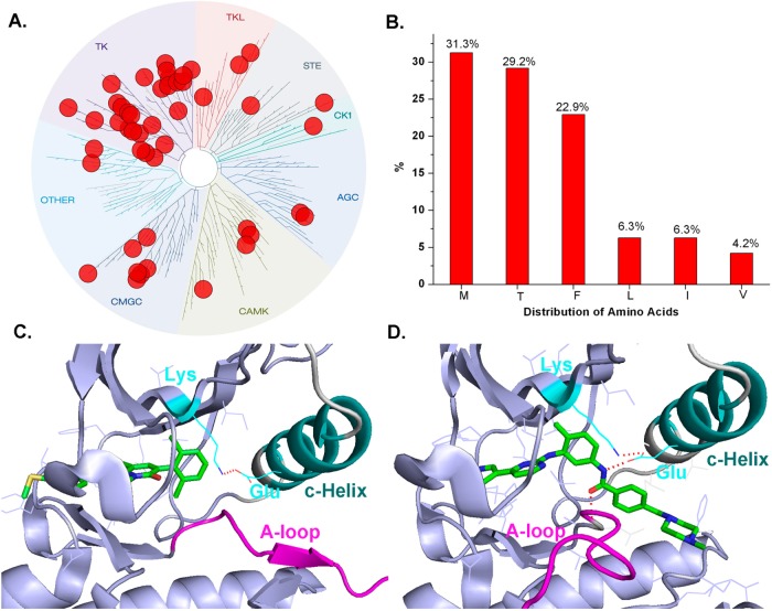
Figure 4.
Analysis of X-ray crystal structure confirmed type II conformation (DFG-out) kinases. (A) Treespot demonstration of type II conformation (DFG-out) kinase distribution. (B) Analysis of gate keeper residues. (C) Demonstration of DFG-out A-loop-R (direct to C-Helix) conformation (PDB ID: 1M52). (D) Demonstration of DFG-out A-loop-L (direct to Hinge) conformation (PDB ID: 1IEP).

