
Figure 2.
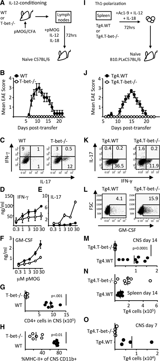
Pathogenicity of Th1 cells is T-bet dependent. (A–H) EAE was induced in C57BL/6 mice by passive transfer of WT or T-bet−/− IL-12-conditioned pMOG-responsive cells. (B) Clinical course of disease is shown. (C) IFN-γ and IL-17-producing capacity of cells on the day of transfer was assessed by flow cytometry. (D–H) Cohorts of mice were sacrificed at 12 days posttransfer to assess cytokine production and CNS inflammation. The production of (D) IFN-γ, (E) IL-17, and (F) GM-CSF by pMOG-stimulated splenocytes is shown. (E) p < 0.001 and (F) p < 0.05, two-way ANOVA with Bonferroni posttest. (G) The numbers of CD4+ T cells in the CNS and (H) the percentage of CNS CD11b+ cells expressing MHC class II in recipients of WT (closed symbols) and T-bet−/− cells (open symbols) are shown. (A–H) All data shown are from one experiment representative of two independent experiments with six mice per group per experiment. (I–O) EAE was induced in B10.PLxC57BL/6 mice by passive transfer of Th1-polarized Tg4.WT or Tg4.T-bet−/− T cells. (J) Clinical course of disease is shown. Results shown are from one experiment representative of three independent experiments with eight to nine mice per group per experiment. Data are shown as mean ± SEM. (K) IFN-γ and IL-17 and (L) GM-CSF producing capacity of cells on the day of transfer are shown. (M–O) The numbers of transferred Tg4 cells retrieved from (M) the CNS and (N) spleen at 14 days posttransfer or (O) the CNS at 7 days posttransfer are shown.