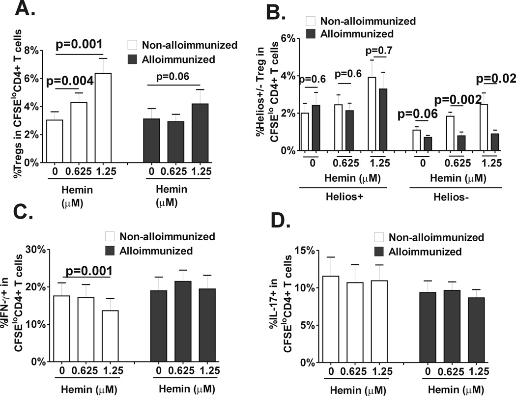
Fig. 3. Differences in Treg/Th polarization in response to hemin between alloimmunized and non-alloimmunized SCD patients.
Purified, CFSE-stained CD4+ T cells from regularly transfused non-alloimmunized (n=9, white bars) and alloimmunized (n=11, black bars) SCD patients were co-cultured with autologous purified total monocyte fraction in the absence or presence of 2 different concentrations of hemin (0.625µM and 1.25µM) and stimulated with anti-CD3 antibody for 7 days. Levels of (A) total Tregs, (B) helios+/− Treg subsets, (C) Th1 and (D) Th17 in CD4+ T population that had undergone proliferation were analyzed by flow cytometry. All statistical analysis comparing before and after hemin addition was performed using paired t test; comparison between alloimmunized and non-alloimmunized groups was performed using Mann-Whitney test.

