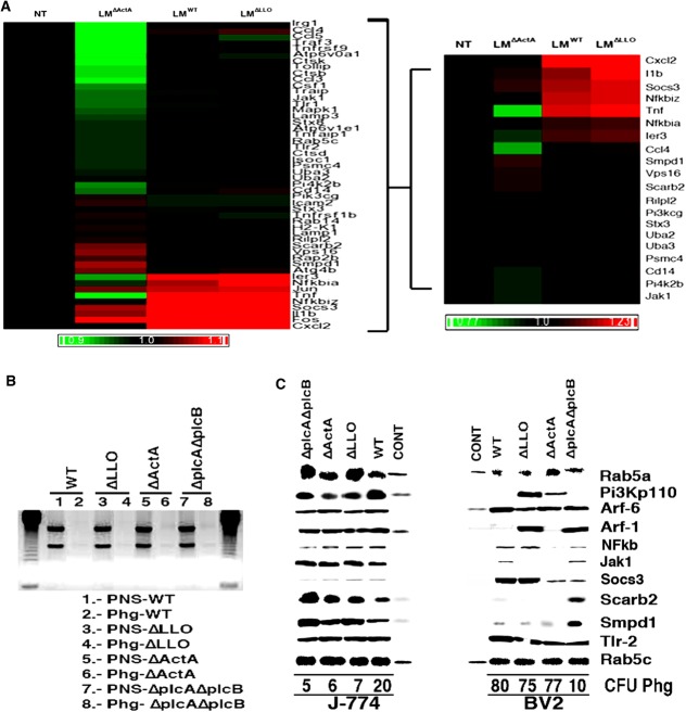
FIGURE 2.
LM actA gene regulates TNF-mediated immune gene expression in microglia, which transforms phagosomes into deficient innate immune platforms. (A) Noninfected (NT) or LM-infected (LMWT, LMΔLLO, or LMΔActA) BV2 cells were used for RNA isolation and differential microarrays. Heat Map presentation of the 20 highest differentially expressed genes representing the LM innate immunity cluster (see Supp. Info. Fig. S3). Colored rows represent expression ratios from ≤ –1.2-fold-change (FC)-repressed genes shown in green to ≥1.2 FC-induced genes shown in red. Black boxes correspond to nondifferentially-expressed genes. (B) Examination of RNA quality in phagosomal preparations. By using 1% agarose gel, RNA major bands, a small ∼2 kb and a large ∼5 kb band, and no fragmented RNA were visualized. In PNSs we observed rRNA (lanes 1, 3, 5, and 7), although no detectable rRNA was observed in phagosomes (lanes 2, 4, 6, and 8). J-774 and BV2 phagosomes usually contain yields of proteins ranging from ∼1 mg/mL and 1–3% of RNA contamination. (C) Protein and functional analysis of phagosomes and endosomes as basal controls. Western blots of 30 µg per lane of J-774 or BV2 isolated phagosomes containing LMWT, LMΔLLO, LMΔActA, or LMΔplcAΔplcB or endosomes from noninfected J-774 or BV2 (CONT lanes) showed different relevant proteins, TLR-2, Pi3kp110, NFkB, Jak1, Socs3, Arf-1, Arf-6, and Rab5a, as well as lysosomal markers, Scarb2, and Smpd1. Rab5c was selected as an internal control marker because it showed no variation in J-774 phagosomes and it is also found in endosomes. CFU numbers under western-blot lanes reflected the amount of live bacteria within the phagosomes. [Color figure can be viewed in the online issue, which is available at http://wileyonlinelibrary.com.]

