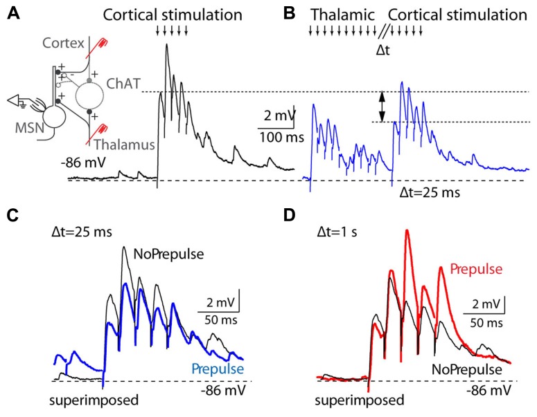
FIGURE 5.
Thalamostriatal projections gate corticostriatal inputs in mouse slices. (A) Left, diagram of the experimental preparation: medium spiny neuron (MSN) recorded in the striatum while corticostriatal projections are activated, with or without preceding stimulation of thalamostriatal projections. Right, activation (downward arrows) of corticostriatal input evokes a train of EPSPs in a MSN cell. (B) Corticostriatal EPSPs are reduced when thalamostriatal stimulation precedes the corticostriatal stimulation by 25 ms. (C) Overlay of corticostriatal EPSPs before and after (blue) thalamostriatal activation to illustrate the changes in amplitude. (D) Overlay of corticostriatal EPSPs before and after (red) thalamostriatal activation, but with a long delay (1 s) between the thalamostriatal and corticostriatal activation. [Reprinted from (Ding et al., 2010), with permission from Elsevier.]

