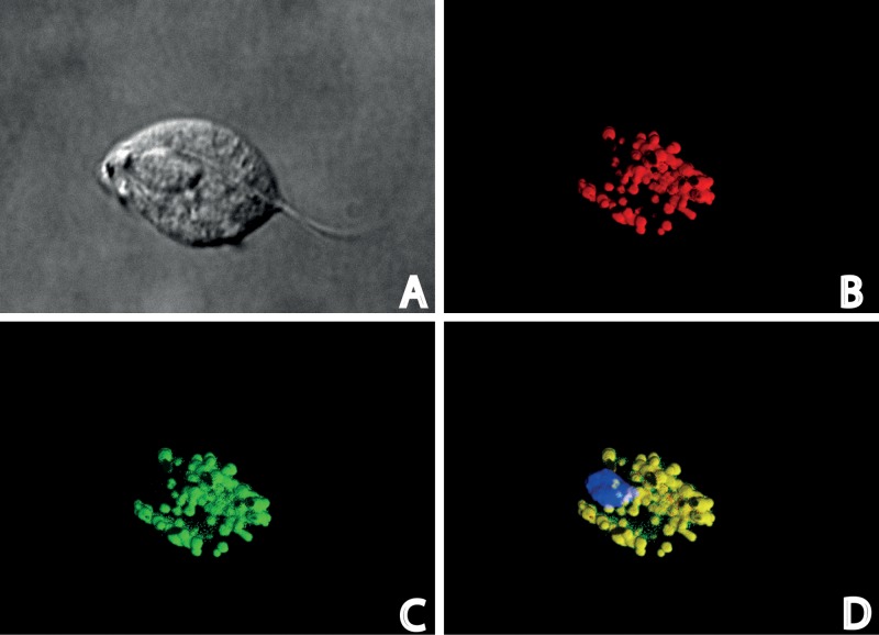
FIG 5.
Immunodetection of TvIsf3 in T. vaginalis cells. (A) Nomarski differential contrast; (B) visualization of malic enzyme, a hydrogenosomal marker; (C) TvIsf3 labeling; (D) merged image of color channels showing the localization of TvIsf3 within the hydrogenosome with DAPI (4′,6′-diamidino-2-phenylindole) staining for nuclei.

