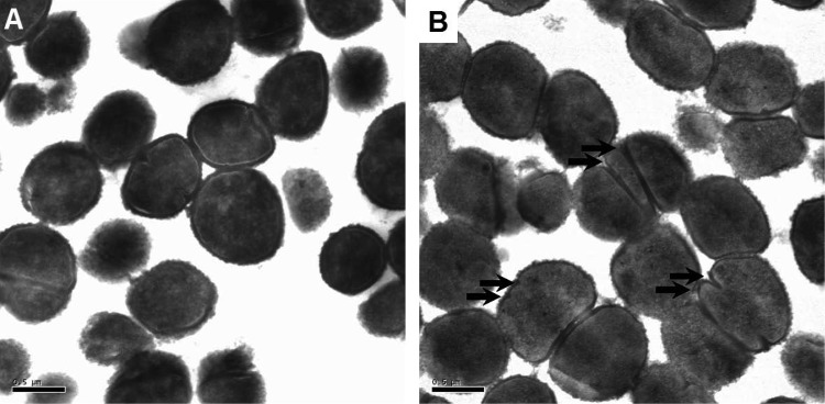
FIG 6.

S. aureus morphology in the presence of C6H. Transmission electron microscopy was used to investigate the morphology of S. aureus after a 2-h standard killing assay in the presence (B) or absence (A) of 5 μg/ml C6H. Scale bars, 0.5 μm. The arrows highlight aberrant septation events.
