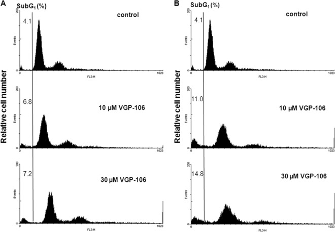
FIG 10.
Effect of VGP-106 on the L. donovani cell cycle. DNA fragmentation was quantified by measuring the percentage of cells in the sub-G1 DNA region. The DNA content degradation profiles of promastigotes were determined by flow cytometry and PI staining. Parasites were incubated without (control) or with 10 and 30 μM VGP-106 for 24 h (A) and 48 h (B) and then loaded with PI, as described in Materials and Methods. The distribution of DNA content was analyzed by flow cytometry. Histograms are representative of three independent experiments, with 10,000 parasites analyzed per group.

