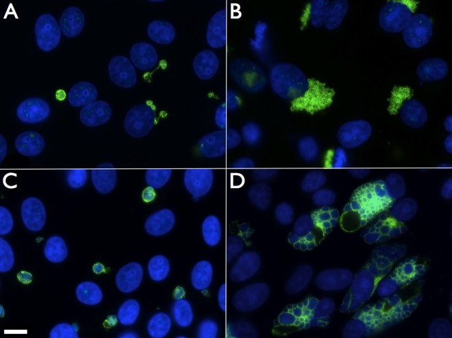
FIG 1.
ST-669 treatment alters the size and structure of C. caviae inclusions. Immunofluorescence microscopy of ST-669 (A and C)- and DMSO (B and D)-treated C. caviae-infected Vero cells fixed at 20 h (A and B) or 40 h (C and D) postinfection was performed. Images were taken at ×100 magnification, and the scale bar in panel C indicates 10 μm for each panel. C. caviae inclusions were labeled with anti-IncA (green), and total DNA was labeled with DAPI (blue).

